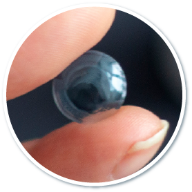
Ocular surface and corneal diseases: cornea conus, iritis

Ocular surface and corneal diseases:
Keratoconus, Iritis and Pterygium

Ocular surface and corneal diseases, such as keratoconus, iritis and pterygium, all affect our eyes in different ways. Keratoconus- can affect the eye’s refractive ability and accelerate the progression of astigmatism. Corneal collagen cross‑linking strengthens the cornea by binding the collagen fibers more tightly together, providing treatment for keratoconus.Iritis, a viral or bacterial infection of the anterior part of the uvea, could lead to complications such as examination before cataract surgery, glaucoma and other complications. And Pterygium is a benign conjunctival lesion, a triangular wing-shaped foreign body that covers the white of the eye and extends to the cornea. When the pterygium gradually grows toward the pupil, it may become inflamed and cause the white of the eye to become red and swollen.It is a benign conjunctival growth, appearing as a triangular, wing‑shaped tissue that spreads over the white of the eye and extends to the cornea. When the pterygium gradually grows toward the pupil, it may become inflamed, causing redness and swelling of the eye. If these symptoms occur, please contact our center promptly to arrange diagnosis and treatment.

Keratoconus

Keratoconus

Also known as keratoconus, this condition occurs when the central cornea becomes thin and is pushed outward by intraocular pressure, altering its normal curvature and causing the cornea to bulge into a cone‑like shape. Keratoconus can impair the eye’s refractive ability and accelerate the progression of astigmatism. The exact cause is still unclear, but it may be related to frequent eye rubbing, allergic diseases, or genetic factors. Symptoms usually appear during puberty or in the twenties. Since early keratoconus resembles common astigmatism or myopia, patients may not realize they have a problem or seek medical advice, making diagnosis more difficult. As the condition progresses, vision may suddenly deteriorate, and even after treatment, corneal scarring can lead to permanent visual impairment.

Symptoms of Keratoconus

Symptoms of Keratoconus: cornea bulges forward

Bulging outward of cornea

Symptoms of Keratoconus: Worsening in a greater and faster manner in myopia or astigmatism

Worsening in a greater and faster manner in myopia or astigmatism

Symptoms of Keratoconus Decreased visual function

Decreased visual function

Treatments of Keratoconus

Management of Keratoconus

Hard contact lens

Hard contact lenses exert greater pressure on the top, so they can press the cornea to deform. However, they are less comfortable and may be displaced on the surface of the eyes.

Keratoconus Treatment Hard Contact Lenses

Corneal collagen cross-linking

Corneal collagen cross-linking surgery (also called corneal cross-linking surgery) is to apply riboflavin (a type of vitamin B2) eye solution to the patient's eyes, and then illuminate the eyes with ultraviolet light for 45 minutes to tighten the collagen in the corneal fibers, strengthening the cornea and making it stronger and more stable. This corneal cross-linking surgery reduces the chance of corneal ectasia, deformation and degree rebound.

  • Avoid eye makeup
  • Avoid getting shampoo or soap into your eyes
  • Avoid getting perfume or hair spray into your eyes
  • Do not drive until your vision is stable
  • No swimming or steam bathing
  • Avoid strenuous exercise, such as health dance and ball games
  • Avoid drinking alcohol and smoking to avoid eye irritation
  • Wear sunglasses when going out to prevent dust or foreign objects from entering your eyes
  • Avoid places with turbid air
  • Follow the doctor's instructions to instill eye drops and follow up regularly.
Corneal collagen cross-linking

Implantation of Intra-Corneal Ring

Intra-Corneal Ring are two crescent-shaped pieces of a plastic that are implanted into the cornea stroma after a tiny incision is made in the cornea. Such procedure is to support the thin and weak cornea and flatten the central curvature of the cornea, which reduces irregular astigmatism and improves visual function. If postoperative vision improvement is not satisfactory, the Intra-Corneal Ring can be replaced or removed.

Keratoconus Treatment Implantation of Intra-Corneal Ring
Iritis

Iritis

The iris is the muscular tissue located between the cornea and the lens. It controls the size of the pupil and forms the front part of the uvea. Therefore, iritis refers to infection of the anterior uvea caused by viruses or bacteria. When the immune system malfunctions and attacks the iris cells, inflammation can also occur, leading to redness of the eye. Without timely treatment, complications such as cataracts or glaucoma may develop. Patients may need oral steroids or anti‑inflammatory eye drops to reduce redness and swelling and relieve eye pain. People often confuse iritis withConjunctivitis. It is often confused with conjunctivitis. However, iritis refers to inflammation inside the eye (the iris), while conjunctivitis refers to inflammation on the surface of the eye (the conjunctiva). Continue reading to learn more about the causes, symptoms, examinations, and management of conjunctivitis.

Symptoms of Iritis

Blurred vision, one of the symptoms of iritis

Blurry vision

Iritis causes sensitivity to light

Sensitivity to light
(photophobia)

Symptoms of Iritis  Eye Pain

Eye pain

Red and itchy eyes, one of the symptoms of iritis

Red and itchy eyes

Iritis causing floaters

Eye floaters

Watery eyes, symptoms of iritis

Watery eyes

Iritis treatment
Steroid drugs

Steroid drugs

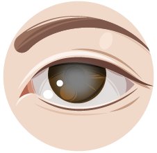
Management of Iritis
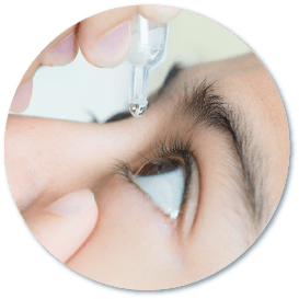
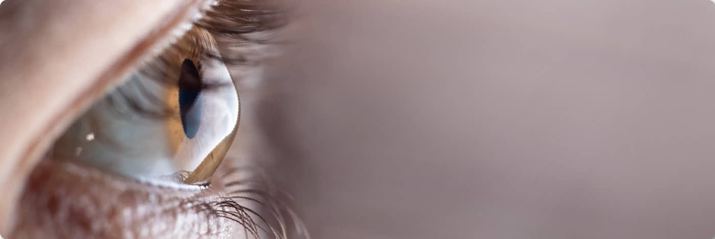
Mild cases

Patients can use steroid eye drops or ointment to control the condition
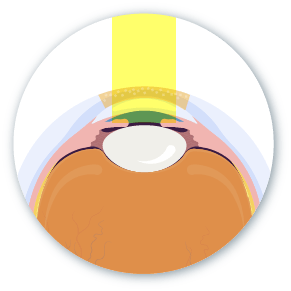
Severe cases

Patients need direct injection of medication into the eye to control inflammation. Some patients even have to take steroids to cure the condition fully.

Caution

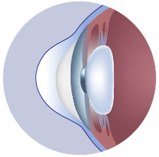
Long-term application of steroids may increase the risk of cataracts and elevated intraocular pressure, so such medications must be prescribed by and under the supervision of an ophthalmologist and should not be purchased on your own.

Pterygium is a benign conjunctival lesion, a triangular wing-shaped foreign body that covers the white of the eye and extends to the cornea. When the pterygium gradually grows toward the pupil, it may become inflamed and cause the white of the eye to become red and swollen.

Pterygium is a benign conjunctival lesion, a triangular wing-shaped foreign body that covers the white of the eye and extends to the cornea. When the pterygium gradually grows toward the pupil, it may become inflamed and cause the white of the eye to become red and swollen.

Pterygium is a benign wedge-shaped fibrovascular growth of the conjunctiva that can enlarge and extend onto the cornea. As the pterygium grows into the pupil, it may become inflamed and cause redness and swelling in the white of the eye. The exact cause of the disease is uncertain. However, prolonged exposure to sunlight or UV light, and irritants like sand and wind will increase the risk of vascular tissue proliferation and increase the chance of pterygium enlargement.

Symptoms of Pterygium

Red and itchy eyes, one of the symptoms of iritis

Red and itchy eyes

Pterygium Foreign body sensation

Foreign body sensation

Pterygium causes visual impairment

Visual impairment

Have burning sensation & inflammation

Burning sensation & inflammation

Some patients have no symptoms

Some patients have no symptoms

Pterygium treatment

Management of Pterygium

Pterygium removal

Until now, there is no medicine that can effectively stop the growth of pterygium, and surgical removal is the only way.

Artificial tears

Artificial tears can moisturise the cornea to relieve dry eyes, control minor symptoms and prevent inflammation.

Please choose to make an appointment or enquiry:
*Please select a clinic
*Enquiry item
We will contact you to confirm the details before confirming the appointment.
*Required
For same day reservation, please call or message us on WhatsApp.