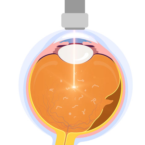
Eye Floaters
Vitreous degradation and opacity due to ageing

What are Eye Floaters?

Do you see any floaters in your field of vision, e.g. small black spots or blotches, threads, fragments of cobwebs? These are all symptoms of eye floaters.

Floaters occur when the normally clear vitreous inside the eye gradually degenerates, becoming liquefied and cloudy. Tiny debris then drifts within the vitreous. When light enters the eye, these cloudy tissues refract the light and cast shadows on the retina, which we perceive as floating spots. Causes include vitreous degeneration, posterior vitreous detachment, and retinal tears. However, if flashes of light or a sudden increase in floaters appear, this may indicate a possible retinal detachment. Immediate medical attention and an eye examination are required, so that an ophthalmologist can provide appropriate treatment to prevent permanent vision loss.

Causes of Eye Floaters

Causes of floaters Vitreous degeneration

Vitreous degeneration due to aging

Aging of the eyes is the most common cause. As age increases, the vitreous body of the eye degenerates and shrinks, gradually detaching from the originally tightly bound retina, and the vitreous body fibers inside may become turbid, projecting many small black dots on the retina, becoming a common sight for people with floaters.

Vitreous detachment from the retinal surface

Vitreous detachment from the retinal surface

Retinal detachment can cause floaters. If floaters are caused by retinal problems, you must seek medical attention immediately, otherwise it may lead to blindness in severe cases.

Causes of floaters holes in retina

Retinal holes

A tear in the retina can cause floaters. If floaters are caused by retinal problems, you must seek medical attention immediately, otherwise it may lead to blindness in severe cases.

Diabetes

Diabetes

Diabetic patients are prone to new and abnormal blood vessels on the retinal surface. The blood vessels are fragile and prone to rupture, making the vitreous hemorrhage and retinal damage prone to blurred vision, making them more susceptible to floaters.

Deep myopia can cause floaters

Deep myopia

The deeper the degree of myopia, the longer and longer the eyeball will become, and the tissue in the eye will be stretched, thinned, and even ruptured, thereby increasing the risk of retinal rupture and detachment, causing floaters.

Eyeball hemorrhage

Eyeball hemorrhage

Eyeball hemorrhage, also known as hemorrhagic floaters, as the name suggests, is caused by retinal hemorrhage, causing dense black spots to appear in front of the eyes, forming floaters.

Causes of floaters Eye inflammation

Eye inflammation

If your eyes have been traumatized or inflamed, impurities may enter the vitreous and cause "floaters" to be seen in your eyes.

Symptoms of Floaters

According to patients' descriptions, common symptoms of floaters include: black spots in front of the eyes, black mesh-like floating objects, flashes of light, and a veil in the field of vision. These symptoms are particularly noticeable in brightly lit areas or against a white background. If the symptoms of floaters increase significantly, you should see an ophthalmologist immediately for a detailed eye examination.

Symptoms of floaters Dark spots

Dark spots

Symptoms of floaters Floating cobwebs

Floating cobwebs

Symptoms of floaters Flashes in vision

Flashes in vision

Common symptoms of floaters Veil crossing vision

Veil crossing vision

Management of Floaters

In most cases, if floaters do not interfere with daily life, no treatment is necessary and patients simply adapt to living with them. However, if the floaters are very large or numerous, appear suddenly, or are accompanied by flashes of light that affect vision, it is recommended to seek prompt consultation with an ophthalmologist for possible surgical or laser treatment. In addition, patients with floaters are advised to undergo a retinal examination every six to twelve months. If symptoms worsen during this period, an earlier follow‑up and comprehensive examination should be arranged.

Laser Treatment

Laser treatment for floaters is mainly used to target larger, more concentrated vitreous opacities, breaking them into smaller fragments to reduce or eliminate symptoms. The procedure is painless and carried out under local anesthesia. However, not all patients are suitable candidates. For example, if the floaters are too diffuse or located too close to the macula or lens, the treatment is not recommended, as it may lead to complications such as cataracts or macular damage.

Floaters laser treatment

Vitrectomy

The vitreous inside the eye is removed through a small incision and replaced with a solution to maintain the eye's shape. Vitrectomy does not necessarily completely remove floaters, and new floaters may form if the surgery itself causes bleeding or retinal tears.

Floaters treatment Vitrectomy

It is crucial to have regular eye examinations so that your doctor can be aware of any changes and help detect eye floaters and other abnormalities.

Floaters FAQ

When the transparent vitreous body in the eye gradually deteriorates, the vitreous body will liquefy and become cloudy, forming small debris floating in the vitreous body. There may also be flashes of light or a sudden large number of floaters due to retinal detachment, so regular eye examinations are required to follow up on the condition of the eyes.

Eye floaters do not go away over time, but they do become less noticeable and settle at the bottom of your eye.

In general, they do not affect the visual acuity and you can get used to floaters. If you see flashes or a sudden appearance of many floaters, these symptoms may indicate retinal detachment, which can be serious enough to cause permanent visual loss.

There are 2 surgical options: 

Laser Treatment

Laser treatment is used for larger and more concentrated vitreous floaters, breaking them into smaller fragments to reduce or eliminate symptoms. However, not everyone is a suitable candidate. For example, if the floaters are more diffuse or located too close to the macula or lens, treatment is not advised, as it may increase the risk of complications such as cataracts or macular damage.
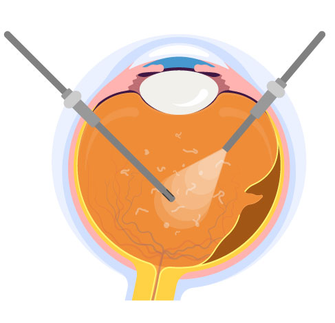
Vitrectomy

The vitreous humour is removed through a small incision and replaced with a solution to maintain the shape of the eye. Vitrectomy does not necessarily remove the floaters completely, and new floaters may form if the surgery itself causes bleeding or slash in the retina.

Physiological floaters:

  • Normal physiological changes in the vitreous body
  • The vitreous body has congenital impurities in it

Pathological floaters:

  • Retinal tears, peeling, punctures, and degeneration
  • Vitreous hemorrhage
  • Macular degeneration
  • Eye bleeding and inflammation caused by diseases such as diabetes, hypertension, and ocular stroke

Diabetic patients are prone to new and abnormal blood vessels on the retinal surface. The blood vessels are fragile and prone to rupture, making the vitreous hemorrhage and retinal damage prone to blurred vision, making them more susceptible to floaters.

Please choose to make an appointment or enquiry:
*Please select a clinic
*Enquiry item
We will contact you to confirm the details before confirming the appointment.
*Required
For same day reservation, please call or message us on WhatsApp.